79 y/o male with Recurrent CVA and left hemiplegia with Aspiration pneumonia and seizures disorder
This is an a online e log book to discuss our patient de-identified health data shared after taking his / her / guardians signed informed consent. Here we discuss our individual patients problems through series of inputs from available global online community of experts with an aim to solve those patients clinical problem with collective current best evident based input.
iv>
>
This E blog also reflects my patient centered online learning portfolio and your valuable inputs on the comment box is welcome.
I have been given this case to solve in an attempt to understand the topic of " patient clinical data analysis" to develop my competency in reading and comprehending clinical data including history, clinical findings, investigations and come up with diagnosis and treatment plan
COMPLAINTS AND DURATION:
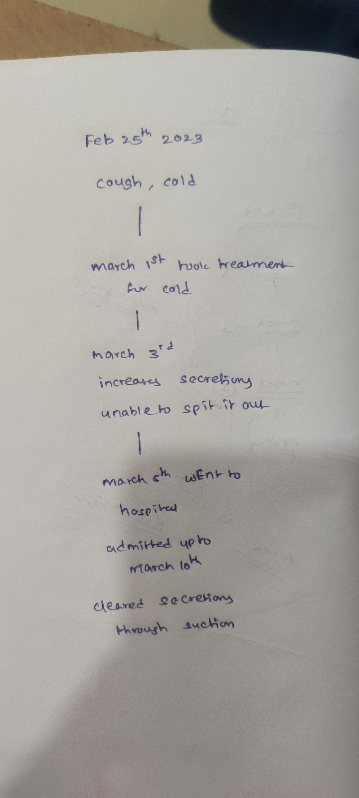
A 79 y/o male was brought to casuality with c/o cough since 20 days ,
fever since 10 days
difficulty in swallowing and h/o Aspiration pneumonia since one month
C/o altered sensorium since 3 days
HOPI:
Patient was apparently asymptomatic 20days back then he developed cough insidious in onset and gradually progressive. PRODUCTIVE but patient is not able to spit it out. Difficulty in swallowing.
H/o cough on intake of liquids.
H/o change of voice since 20 days, insidious, hoarse in character and
SLURRING OF SPEECH +present
No h/o difficulty in breathing, breathlessness, hemoptysis
Fever since 10 days -high grade. O/e Chills and rigors + (38 spikes).
N/h/o vomiting, chest pain, loose stools.
7 YEARS BACK:(2016)
He developed head ache at around afternon 2pm and followed by vomtings and left hand itching and weakness.
PATIENT is awake on that night due to left hand weakness and itching
NEXT DAY
MORNING they took him to hospital
Patient can lift his hand
But unable to hold objects
AFTER 3 DAYS
PATIENT became left sided hemiplegia.
MRI REPORT- 3 INFARCTS
Patient stayed for 40 days in hospital and there was no improvement and discharged.
He took liquids for 3months because patient is unable to eat solid foods.then he slowing started eating solid foods.
AFTER 1 YEAR (2017):
vomitings
Fever
Shivering for 3 days
Diagnosed with urinary tract infection
Took treatment (antibiotics) for 5 days and it resolved
AFTER 3 YEARS:(2020)
Cough for 2days
Fever on 2nd day
Diagnosed with covid
He got COVID for 1st time and resolved
After 1 year(2021)
He was Diagnosed with COVID for 2nd time and resolved
1 YEARS back (2022)
He got seizures for 5min and they took him to the hospital.
He got Typhoid fever 2times
1st time resolved in 7days
2nd time resolved in 9 days
79 Year old male who is a father of 4 children ( 2 sons and 2 daughters)..was used to run shop ( kirana shop) for about 18 years.He stopped looking after his shop from 2006 and he was looked after by his son's.
He was non alcoholic,non smoker.
10 years back , patient developed lesions on his both foot and went to the doctor and found to have diabetes and started on medication.and after 1 year ,with regular check up he was found to be Hypertensive and started on antihypertensive medication.
From 7 years onwards , patient was bedridden with foleys ( changed every 15 days ) and physiotherapy was done by his attenders daily, but there was no such improvement
PAST HISTORY
Patient is a k/c/o Hypertension and type 2 diabetes since past 10years for which he is on medications I.e tab TELMA AM 40mg po/od. Tab zoryl mv , po/od
PERSONAL HISTORY
Appetite lost,
Mixed diet
Bowel- constipated,
Bladder regular
No known allergies and Addictions.
i.e non alcoholic and non smoker
Family History- not any
Treatment history
•Tab TELMA AM 40mg po/od since past 10years
•Tab zoryl mv , po/od
•Tab levipil 500mg since 2 years
• thyronorm 25mcg. Since5 years
GENERAL EXAMINATION
On examination patient is arousable but not oriented.
Pt not cooperative mostly.
-PALLOR: PRESENT
-NO PEDAL EDEMA, ICTERUS, CYANOSIS, CLUBBING, LYMPHADENOPATHY
VITALS ON ADMISSION
PR-90 BPM
BP- 140/80MM HG
RR- 22 CPM
SPO2- 98% AT RA
GRBS - 183mg/dl
SYSTEMIC EXAMINATION:
Respiratory :-
Inspection : respiratory movements equal on both sides
Trachea central
palpation : apical impulse in left 5th intercostal space
Auscultation : normal vesicular breath sounds
Percussion- BAE+
CNS
PATIENT is unconscious incoherent uncooperative
HIGHER MENTAL FUNCTIONS- cannot be elecited
Speech
Behaviour
Memory
Intelligence
Lobar functions
B/L PUPILS - NORMAL SIZE AND REACTIVE TO LIGHT
NO SIGNS OF MENINGEAL IRRITATION,
CRANIAL NERVES
2nd cranial nerve
Visual acuity is decreased on left side
3rd 4th 6th pupillary reflex present
SENSORY SYSTEM- cannot be elicited
Spinothalamic sensation:
Crude touch
Pain
Temperature
Dorsal column sensation
Fine touch
Vibration
Propioception
Cortical sensation
Two point discrimination
Tactile localisation
Stereognosis
Graphathesia
MOTOR EXAMINATION:
Right n left
UL. LL. UL. LL
BULKv Normal Normal Reduced
TONE Normal Hypotonia
POWER Could not be elicited
SUPERFICIAL REFLEXS
plantar reflex
Left side babinski sign positive
iv>
• DEEP REFLEXES:
BICEPS, TRICEPS, SUPINATOR, KNEE ANKLE -
>
CEREBELLAR EXAMINATION cannot be elicited
Finger nose test
Heel knee test
Dysdiadochokinesia
Dysmetria
hypotonia with pendular knee jerk present.
Intention tremor present.
Rebound phenomenon .
Nystagmus
Titubation
Speech
Rhombergs test
SIGNS OF MENINGEAL IRRITATION: absent
GAIT: patient unable to walk
CVS
ASCULTATION: S1S2 +,NO MURMURS
P/A
INSPECTION: UMBILICUS IS CENTRAL AND INVERTED, ALL QUADRANTS MOVING EQUALLY WITH RESPIRATION,NO SCARS,SINUSES, ENGORGED VEINS, PULSATIONS
AUSCULTATION: no bowel sounds heard
bed sores
C/o asymptomatic lesions all over the body since 2 months
H/o application of unknown topical medications used
O/e multiple hyperpigmented Macclesfield present all over the body with scaly lesions over the upper back
•Diffuse xerosispresent
• single ulcer of size 1.5x1.5 cms (approx) over the back.
Diagnosis SENILE XEROSIS + post inflammatory hyperpigmentation.
( +? TROPHIC ULCER )OBSERVATIONS:
• Large area of encephaolomalacia in right occipito -temporo lobes and righ parietal lobes.
• Prominence of sulci and cisterns.
• Bilateral periventricular hyperintensity.
• Rest of the Cerebral parenchyma shows normal gray/white matter differentiation.
• Basal ganglia and Thalami are normal.
• Brain stem normal.
• Cranio-vertebral and Cervico-medullary junctions are normal.
• Sella, pituitary and parasellar regions are normal. Stalk and hypothalamus are normal. Posterior pituitary bright spot is normal.
• No evidence of abnormal calcifications, vascular anomalies on SWI sequences.
IMPRESSION:
• Large area of encephalomalacia in right occipito-temporo lobes and right parietal lobes - sequelae of old infarct.
• Diffuse cerebral atrophy. Chronic small vessel ischemia.
Note: Poor quality of images due to motion artefacts
CUE :-
AFB-TRACE
PUS CELLS -2-4
EPITHELIAL CELLS -2-3
LFT
INVESTIGATIONS:
Anti HCV antibodies rapid -nonreactive
Blood urea -30mg/dl
HBA1C-6.7%
HbsAg rapid - negative
HIV 1/2 RAPID TEST - NON REACTIVE
TOTAL BILIRUBIN -0.81mg/dl(normal-0 to 1mg/dl)
Direct bilirubin-0.17mg/dl(0 to 0.2mg /dl)
Serum creatinine -0.9 mg/dl (0.8 to 1.3 mg /dl)
ABG
Ph 7.51
PCO2 29.5mmhg
Po2 67.5 mmhg
Electrolyte
Sodium 135meq/l
Potassium 3.5 meq/l
Chloride 98meq/l
Calcium -1.06 mmol/l
PROVISIONAL DIAGNOSIS
Recurrent CVA with Hypertension, T2 DM, seizures disorder.
TREATMENT
1) TAB ECOSPRIN 150 mg RT/OD
2) TAB CLOPIDOGREL 75 MG RT/OD
3) TAB ATORVAS 20 MG RT/OD
4) NEBULISATION - 3% NS ,
MUCUMZY 8th hourly
5) CHEST PHYSIOTHERAPY.
6) RT FEEDS 100 ML WATER 2nd HRLY
50 ML Milk 2nd HRLY.
8) TAB. THYRONORM 25MCG RT/OD
9) TAB. LEVIPiL


Comments
Post a Comment